
Introduction
Mycoplasma pneumoniae is a frequent cause of community-acquired pneumonia, accounting for approximately 10% of cases and up to 37% of pediatric cases.1,2 M pneumoniae is classified as an atypical bacterium due to its lack of a cell wall. It is inherently resistant to beta-lactam antibiotics, including first-line agents such as penicillins and cephalosporins, commonly used to treat bacterial pneumonia.3 It leads to extrapulmonary complications in about 25% of patients, including Mycoplasma pneumoniae-induced rash and mucositis (MIRM), a distinct condition marked by painful mucositis affecting the oral, ocular, and urogenital mucosae with minimal or absent skin involvement.1,4 Predominantly affecting children and adolescents (mean age ~12 years, male predominance), MIRM presents with erosive or ulcerative mucosal lesions and, when present, sparse vesiculobullous or targetoid skin lesions covering less than 10% of the body surface area.5,6 Its clinical overlap with Stevens-Johnson syndrome (SJS), erythema multiforme (EM), and herpes simplex virus (HSV) infections often leads to misdiagnosis, as seen in this case of a six-year-old female initially treated for HSV due to mucosal ulcerations.5 There have also been other cases of common pathogens, for example, Chlamydophila pneumoniae, that presented with rare symptoms of mucocutaneous disease similar to MIRM.7 This report highlights MIRM’s unique features, the diagnostic challenges in distinguishing it from HSV and other mimics, and the importance of early recognition to ensure appropriate management and prevent unnecessary treatments. To the best of our knowledge, this is the first report that emphasizes the importance of considering MIRM in the differential diagnosis of patients presenting with clinical HSV cutaneous manifestations.
Case Report
A six-year-old female presented to her primary care physician (PCP) for her annual physical, reporting acute left-sided ear pain (otalgia) of two days’ duration. Physical examination revealed purulent otorrhea in the left ear canal, with partial obstruction of the tympanic membrane, alongside mild nasal congestion. She was diagnosed with acute otitis media and prescribed Augmentin (amoxicillin-clavulanate) 875 mg twice daily, Ciprodex (ciprofloxacin-dexamethasone) otic drops, and Ofloxacin otic solution. After 14 days, the otalgia persisted, now accompanied by new-onset throat pain and lip discomfort described as burning. Examination showed erythematous pharyngeal mucosa and mild lip swelling; she was prescribed Cefadroxil 500 mg twice daily for presumed pharyngitis.
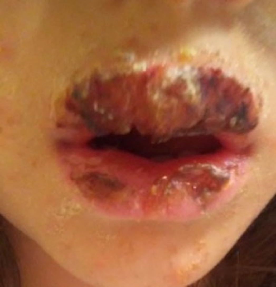
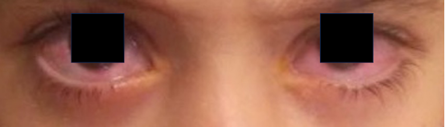
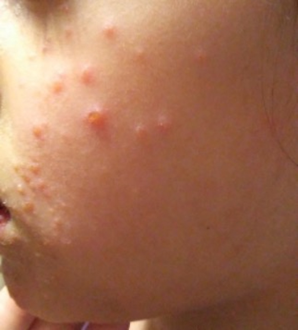
On day 24, during a telehealth visit, the patient exhibited a mild papular dermatitis on her abdomen, elbows, and face, characterized by scattered erythematous papules (Figure 1). Suspecting herpes simplex virus (HSV) infection due to the distribution and mucosal involvement, the PCP prescribed topical Abreva (docosanol). Over the next four days, her condition worsened significantly: the lip pain intensified, evolving into painful ulcerative mucositis with hemorrhagic crusting (Figure 2), and she developed bilateral ocular redness, photophobia, and periorbital edema suggestive of conjunctivitis (Figure 3). Acyclovir (20 mg/kg orally four times daily) was initiated for presumed HSV mucocutaneous disease. The PCP referred her to an ophthalmologist, who noted bilateral lid and conjunctival injection and mucoid discharge, reinforcing a diagnosis of herpes conjunctivitis.


Despite antiviral therapy, symptoms progressed over the next three days, with worsening mucositis and ocular involvement prompting referral to a dermatologist. Physical exam revealed extensive oral ulcers with sloughing mucosa, scattered papules with central vesiculation on the trunk and extremities (covering <5% body surface area), and no epidermal detachment. Given the lack of improvement with antivirals and atypical features (minimal cutaneous spread, prominent mucositis), serological testing was ordered. Results returned positive for Mycoplasma pneumoniae IgM and IgG with negative HSV PCR and cultures, confirming Mycoplasma pneumoniae infection (Table 1). Combined with her clinical presentation, a diagnosis of Mycoplasma pneumoniae-induced rash and mucositis (MIRM) was established.
Treatment was promptly adjusted to Azithromycin (10 mg/kg on day 1, then 5 mg/kg daily for 4 days) and topical Clindamycin for secondary bacterial coverage of mucosal lesions. Within 48 hours, her ocular symptoms began to resolve, and by day 7 of antibiotic therapy, the mucositis and dermatitis significantly improved, with near-complete resolution by day 14. Follow-up at one month showed no recurrence, though mild lip dryness persisted.
Discussion
In this report, we note the difficulty of diagnosing MIRM in pediatric patients. MIRM’s mucosal-predominant presentation, with minimal cutaneous involvement (<5% body surface area in this case), overlaps with Stevens-Johnson syndrome (SJS), toxic epidermal necrolysis (TEN), erythema multiforme (EM), and HSV, often leading to misdiagnosis.1,5,6 The mucosal lesions are often erosive or ulcerative and may be accompanied by conjunctivitis or genital ulcerations, for example, Vujic et al. (2015) described a young adult with severe oral and ocular mucositis but no skin involvement, initially mistaken for SJS until serologic evidence confirmed MIRM.8,9 Similarly, Poddighe et al. (2017) reported a pediatric patient with oral, genital, and anal mucositis, initially evaluated for HSV and Behçet’s disease before M. pneumoniae was identified.10 These cases underscore the need for diagnostic vigilance in mucosal predominant presentations.
To distinguish MIRM, Canavan et al. (2015) propose diagnostic criteria: clinical or laboratory evidence of M. pneumoniae infection (e.g., positive IgM/IgG serology, PCR), involvement of at least two mucosal sites (94% oral, 82% ocular, 63% urogenital), and sparse or absent cutaneous lesions (<10% body surface area, 34% with no skin involvement).5 Unlike SJS/TEN, which are typically drug-induced (e.g., antibiotics, antiepileptics) with extensive epidermal detachment, MIRM follows a respiratory prodrome (fever, cough, malaise ~1 week prior), as seen in our patient with initial otalgia and congestion.11 EM, often HSV associated, features prominent targetoid skin lesions, whereas MIRM’s skin lesions, if present, are vesiculobullous or atypical targetoid.5,6 Santos et al. (2020) further note that MIRM’s milder prognosis (81% full recovery, 3% mortality) contrasts with SJS/TEN’s severe morbidity, and its lack of HSV PCR positivity helps rule out viral mimics.12 In our case, negative HSV PCR and positive M. pneumoniae IgM/IgG (titers 1:128/1:256) were pivotal in correcting the initial HSV misdiagnosis.
Rapid diagnosis is critical to avoid inappropriate treatments, as seen with the antiviral therapy (acyclovir) prescribed here, which delayed effective management. Early M. pneumoniae testing with serologic assays (IgM/IgG), PCR, or cold agglutinin titers, in young patients with mucosal predominant symptoms and a recent respiratory illness is recommended.5 A structured diagnostic approach, including a detailed history for respiratory prodrome, prompt serologic/PCR testing, and exclusion of HSV or cytomegalovirus via PCR is imperative to streamline diagnosis of MIRM.12 In atypical cases, consultation with infectious disease or dermatology specialists can expedite accurate identification, as demonstrated by the dermatologist’s role in our case.
Management involved azithromycin, effective against M. pneumoniae’s beta-lactam resistance, and supportive care, leading to near complete resolution within 14 days.3 This aligns with MIRM’s favorable prognosis when correctly identified.5 However, misdiagnosis, as seen in historical cases mistaken for SJS or EM, can lead to unnecessary antivirals, ineffective antibiotics, or immunosuppression, prolonging morbidity.9,10 This case underscores the importance of including MIRM in the differential for pediatric mucosal eruptions, particularly when HSV is suspected, and prioritizing early M. pneumoniae testing to ensure timely treatment and minimize complications.
Conclusion
This case report seeks to add to the limited literature on this topic by describing the diagnosis and management of a patient with MIRM and enhancing the differential diagnosis for pediatric skin conditions when HSV is suspected. The diagnosis of HSV was initially made based on the clinical presentation. Treatment options for HSV include antiviral medications, while MIRM is usually treated with antibiotics. Differentiating between these two conditions is crucial, as treatment approaches differ significantly. This case report emphasizes the significance of recognizing the difference between MIRM and HSV infection in pediatric patients and emphasizes the importance of prompt and accurate diagnosis to avoid unnecessary suffering and complications.